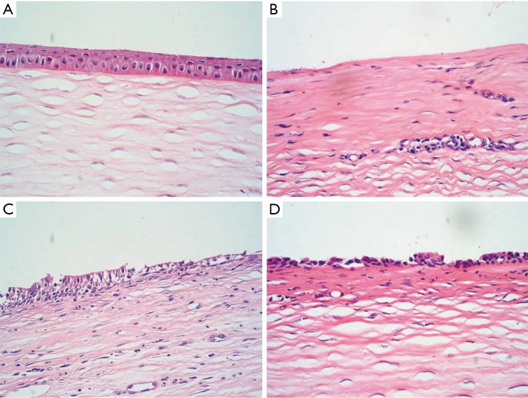
Figure 6.
Assessment of corneal epithelium repair by H&E staining (×100). (A) Normal corneal epithelium; (B) corneal epithelial defect. There was infiltration of inflammatory cells; (C) loose cells in the superficial layer. A large number of inflammatory cells were present; (D) cells arranged in one or two layers on the surface.

