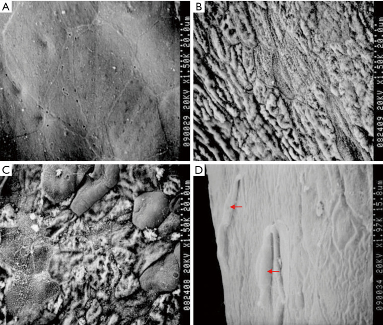
Figure 8.
Observation of the corneal surface using scanning electron microscopy. (A) normal corneal epithelium; (B) the LSCD group. Only exposed collagen fibers are visible; (C) the amniotic membrane transplantation group; (D) the rADSCs transplantation group. Spindle-shaped cells are present (indicated by arrows). LSCD, limbal stem cell deficiency; rADSC, rabbit adipose tissue-derived stem cell.

