Figure 1.

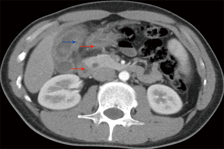
The preoperative CT transverse section proves diffuse asymmetrical gallbladder wall thickening (blue arrow) and contiguous xanthogranuloma lesions invading into adjacent tissues (red arrow).

The preoperative CT transverse section proves diffuse asymmetrical gallbladder wall thickening (blue arrow) and contiguous xanthogranuloma lesions invading into adjacent tissues (red arrow).