Figure 1.
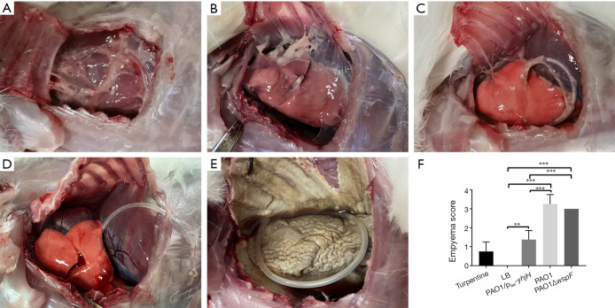
The gross pathology specimens of rabbit pleural cavities 96 h after injection. For PAO1 group (A), PAO1ΔwspF group (B), and PAO1/plac-yhjH group (C), there were varying degrees of pleural adhesions and fibrin depositions between the visceral pleura and parietal pleura. (D) In the LB control group, there were no significant changes in the right pleural cavity. (E) In the turpentine control group, there was severe aseptic inflammation. (F) The empyema score of each group. Results displaying the mean ± SD. An asterisk indicates significant difference, **, P<0.01; ***, P<0.001. The ANOVA tested. (300×300 DPI). LB, Luria-Bertani.

