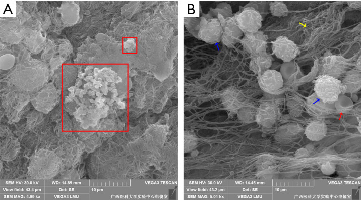
Figure 6.

Observation of the pleural surface by scanning electron microscopy. (A) Biofilm-like structures on the pleural surface (5,000×). (B) Pleural surface host cells (5,000×). PAO1 wild-type strains were embedded in electron-dense extracellular matrix (red box), which appeared to be biofilm-like structures. The blue arrow indicates the polymorphonuclear leukocytes, the yellow arrow indicates the fibrin and the red arrow indicates erythrocyte (300×300 DPI).
