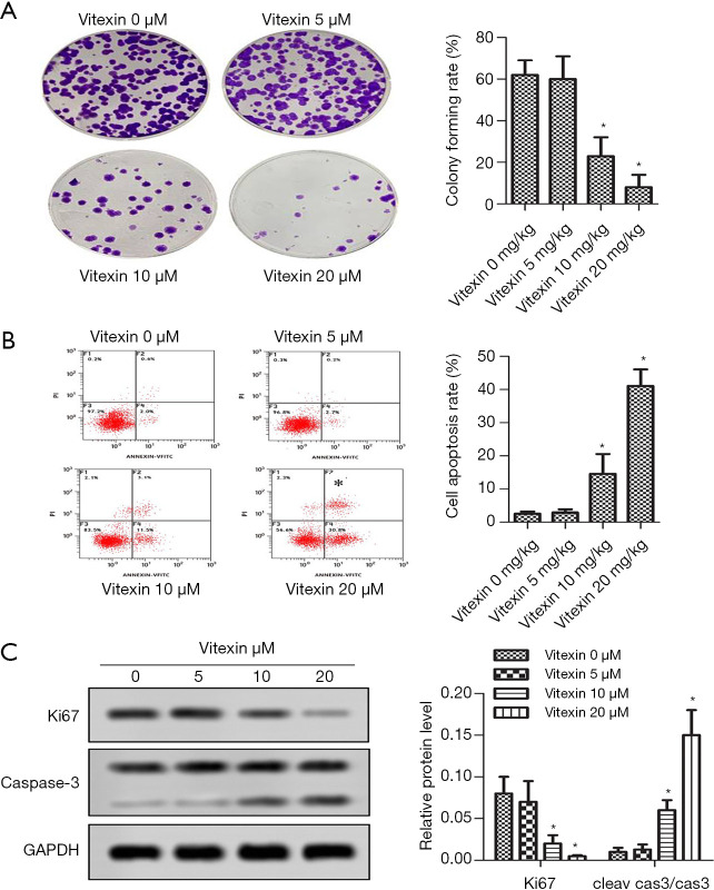
Figure 2.
Vitexin treatment inhibited proliferation and promoted apoptosis of SKOV-3 cells. (A) Proliferation was detected by clonal formation assay (magnification ×200). (B) Cell apoptosis rate was detected by flow cytometry [propidine iodide (PI) staining]. (C) Relative protein levels of Ki67 and caspase-3 were detected by western blot assay (*, P<0.05 vs. control).

