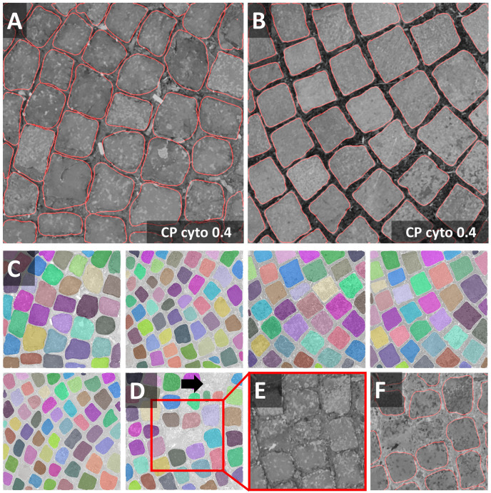
FIGURE 3.
Cobblestones notebook: Segmentation of non-roundish cells (A,B) Segmentations (red line) of the Cellpose Cyto 0.4 model are superimposed onto the original image subjected to [median 3 × 3] preprocessing. The inverted image (not shown) was used as input to the segmentation. Outlines are well defined, no objects were missed, none over-segmented. These settings fit accurately to the entire dataset (train and test) shown in panels (C,D). Only in one image, three objects were missed and one was over-segmented. Borders around these stones are hard to discern. Individual objects are false color-coded in panels (C,D). The red squares in panel (D) highlight one of the two problematic regions shown as a close-up in panels (E,F).

