Figure 7.
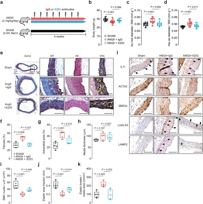
Administration of IL11 antibodies prevents ANGII-induced aortic remodeling. (a) Schematic diagram depicting ANGII experimental protocol. Wildtype C57BL/6 J mice were injected with X203 or IgG control antibodies (20 mg/kg via IP twice per week) starting from 24 h after being implanted with osmotic minipumps containing ANGII. (b) Body weights and aortic echocardiography for (c) aortic root diameter and (d) ascending aortic diameter in Sham, ANGII + IgG, ANGII + X203 animals (n = 11–17/group). (e) Representative photomicrographs captured at 100X and 400X magnification of transverse aortic sections in sham, ANGII + IgG, and ANGII + X203 mice stained with Masson’s Trichrome (MT), hematoxylin and eosin (HE) and Verhoeff Van Gieson (VVG) stains. The white and yellow double-headed arrow demarcates the tunica adventitia and media layers respectively. Black arrows indicate cell-free foci of proteoglycan-rich matrix, white and yellow arrowheads indicate replacement fibrosis in the tunica media and lamellar elastin breaks respectively. Histological analyses of (f) fibrosis*, (g) adventitial area, (h) media thickness, (i) SMC nuclei, (j) elastin area and (k) elastic lamella breaks (n = 5/group). Statistical analyses by one-way ANOVA with Sidak multiple comparisons, unless data deviated significantly from normal, in which case a Kruskal–Wallis test with Dunn’s multiple comparisons was performed (denoted by *). Data presented as median ± IQR, whiskers define the minimum and maximum values. (l) Representative photomicrographs captured at 400X magnification of transverse aortic sections in sham, ANGII + IgG, and ANGII + X203 mice immunostained with anti-IL11, ACTA2, SM22α, LGALS3, and LAMP2 (n = 3/group). Black arrows indicate labeled VSMCs within the media. Scale bars represent 100 µm.

