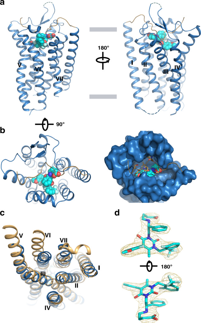
Fig. 1. Overall structure of human GnRH1R in complex with antagonist elagolix.

a View from within the plane of membrane. GnRH1R is shown in sky blue cartoon representation. The N terminus is displayed as sand cartoon. The antagonist elagolix is shown as sphere with cyan carbons. b View from the extracellular side of the membrane. Left one represents sky blue cartoon receptor with cyan sphere ligand. Right one shows the surface representation of 7-TM domain, key residues of the N terminus that are shown in sand sticks inserted the orthosteric pocket. The N terminus is displayed as sand cartoon. c View from the intracellular side of the membrane, structural comparison of GnRH1R (sky blue) with G protein -bound active NTS1R (6OS9, orange). d |Fo| − |Fc| omit map (contoured at 3.0σ) of the ligand elagolix. The antagonist elagolix is shown as cyan sticks.
