Figure 5.
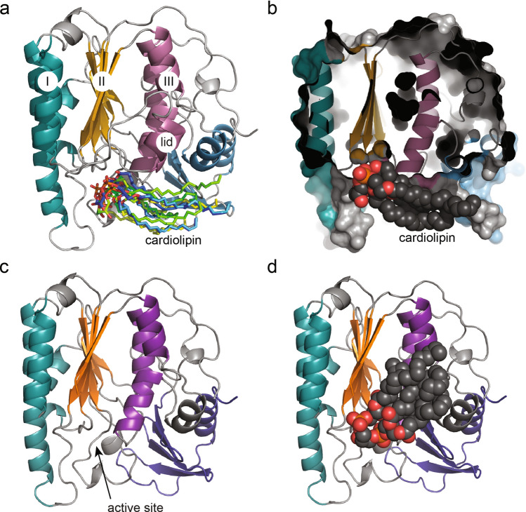
Cardiolipin docking to YejMPD with and without lid. (a) YejMPD structure indicating layer I, II and III, intact lid, and superimposed cardiolipin molecules in their preferred docked location and orientations, with phospholipid heads groups pointing towards the active site and acyl chains towards the CT domain. (b) YejMPD shown in cartoon and surface volume representation cropped in the Z-plane to visualize the vestibule of the active site, Black arrow point towards preferred location of cardiolipin molecule. Gray arrow points towards the hydrophobic pocket and proposed cardiolipin binding site between layers II and III. (c) YejMPD with removed lid, black arrow pointing towards the exposed area of the hydrophobic pocket between layers II and III. (d) YejMPD with removed lid shows cardiolipin molecule with preferred acyl chains flipped upwards towards the hydrophobic pocket while the phospholipid headgroups remain located towards the active site vestibule at the base of layers II and III.

