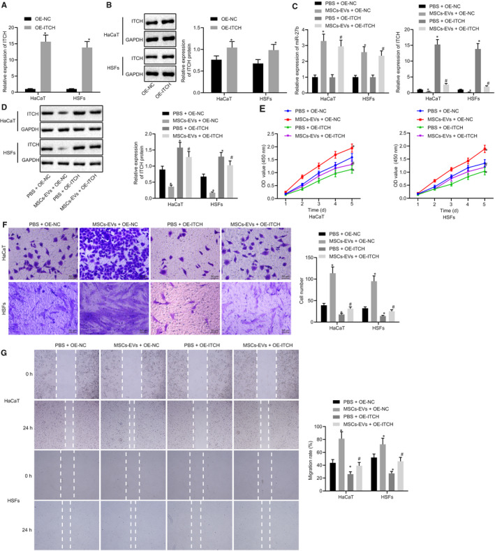
FIGURE 5.

Transfer of miR‐27b via MSC‐derived EVs improves proliferation and migration of HaCaT cells and HSFs by repressing ITCH in vitro. (A) mRNA expression of ITCH was examined by RT‐qPCR in HaCaT cells and HSFs after treatments with OE‐ITCH and OE‐NC, normalized to GAPDH. (B) Representative Western blots of ITCH protein and its quantitation in HaCaT cells and HSFs after treatments with OE‐ITCH and OE‐NC, normalized to GAPDH; *P < .05 compared with OE‐NC treatment. The HaCaT cells and HSFs transfected with OE‐NC and OE‐ITCH used for following detections were treated with PBS and MSC‐derived EVs, respectively. (C) Expression of miR‐27b and ITCH was examined by RT‐qPCR in HaCaT cells and HSFs after different treatments, normalized to U6 and GAPDH, respectively. (D) Representative Western blots of ITCH protein and its quantitation in HaCaT cells and HSFs after different treatments, normalized to GAPDH. (E) Proliferation of HaCaT cells and HSFs was examined by CCK‐8 assay after different treatments. (F) Migration of HaCaT cells and HSFs was examined by Transwell assay after different treatments (scale bar = 50 μm). (G) Migration rate of HaCaT cells and HSFs was determined by scratch test after different treatments. *P < .05 compared with HaCaT cells and HSFs transfected with OE‐NC and co‐treated with PBS; #P < .05 compared with HaCaT cells and HSFs transfected with OE‐NC and co‐treated with MSC‐derived EVs. Data between the two groups were compared by unpaired t test. Data are shown as mean ± standard deviation of three technical replicates
