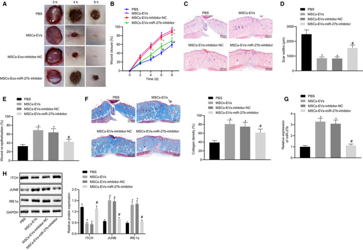
FIGURE 7.

MSC‐derived EVs mitigate epidermal re‐epithelialization and collagen fibre proliferation in wounds by delivering miR‐27b in vivo. The mice used for following detections were treated with PBS, EVs derived from MSCs transfected with inhibitor‐NC or EVs derived from MSCs transfected with miR‐27b‐inhibitor. (A) Size of wounds in mice at the 0, 2, 4, 6 and 8 d after operation. (B) Healing rate of wounds. (C) Pathological changes of cutaneous wound tissues were observed by HE staining in mice at 8 d after operation (scale bar = 250 μm). (D) Quantified scar width in the wound of mice. (E) Wound re‐epithelialization in mice. (F) Masson‐stained collagen in wound tissues of mice (scale bar = 250 μm). (G) miR‐27b expression was determined by RT‐qPCR in cutaneous wound tissues of mice at the 8 d after operation, normalized to U6. (H) Representative Western blots of ITCH, JUNB and IRE1α proteins and their quantitation in cutaneous wound tissues of mice, normalized to GAPDH. *P < .05 compared with PBS treatment; #P < .05 compared with EVs derived from MSCs transfected with inhibitor‐NC by unpaired t test. The data above were measurement data presented as mean ± standard deviation. n = 15 for mice following each treatment
