
Figure 4.
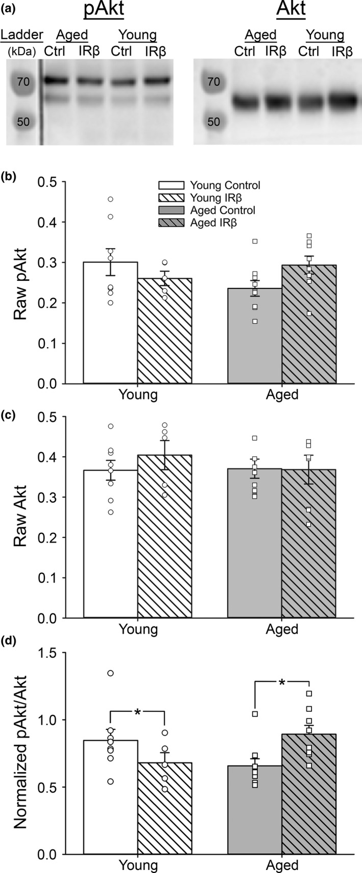
Western immunoblots probing for downstream IR signaling markers in hippocampal tissue from control and IRβ‐treated animals. (a) Representative photomicrograph of Western immunoblots performed on hippocampal tissue taken from 30 animals (young control = 8, young IRβ n = 5, aged control n = 9, aged IRβ n = 8) probing for pAkt (left) or Akt (right). (b‐c) Quantification of pAkt (b) and Akt (c) expression. No differences across age or treatment were detected for either marker (2‐way ANOVA; p > 0.05). (d) Quantification of pAkt/Akt ratios indicate that our manipulation was able to impact downstream IR signaling in the hippocampus, as indicated by a significant interaction (2‐way ANOVA; F (1,26) = 8.09, p = 0.009). Only aged animals responded with increased IR signaling following IRβ treatment. All data were normalized to total protein levels (Ponceau S staining). Asterisks (*) indicate significance at p < 0.05. Data represent means ± SEM