FIGURE 6.

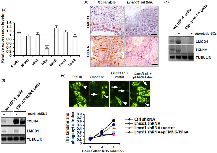
Lmcd1 depletion compromises TXLNA‐mediated testicular phagocytosis. (a) Expression levels of different key factors essential for testicular phagocytosis in Lmcd1 siRNA‐treated testis were determined using RT‐qPCR. **p < 0.01 compared with the values in Ctrl siRNA‐treated testis. (b) Expression levels of LMCD1 and TXLNA in Lmcd1 siRNA‐treated testis were evaluated using immunohistochemistry at translational level. Scale bar, 20 μm. (c) Wild‐type 15P‐1 and 15P‐1Lmcd1−/− cells were incubated with apoptotic GCs for 6 h, followed by immunoblotting analysis. (d) Wild‐type 15P‐1 and 15P‐1/TXLNA cells were transduced with Lmcd1 shRNA for 48 h, followed by immunoblotting analysis. (e) Wild‐type 15P‐1 and 15P‐1Lmcd1−/− cells were transiently transfected with pCMV6‐Txlna or empty vectors. 48 h later, cells were subjected to the RB binding and phagocytosis assay, as described above. **p < 0.01 comparing Lmcd1 shRNA+vector to Lmcd1 shRNA+pCMV6‐Txlna
