FIGURE 1.

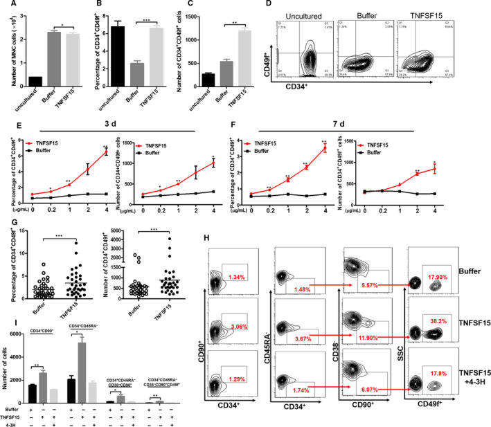
TNFSF15 promotes in vitro expansion of primitive human CD34+CD49f+ haematopoietic stem cells. A, Number of total mononuclear cells after being treated with TNFSF15 for 7 d at 2 µg/mL in expansion medium (n = 3). B, Percentage of CD34+CD49f+ cells after being treated with TNFSF15 for 7 d at 2 µg/mL in expansion medium on the same human umbilical cord blood sample (n = 3). 1 × 10⁴ CD34+ human UCB cells were seeded in the beginning. The experiment was repeated for three times. C, Absolute number and representative pictures (D) of CD34+CD49f+ cells after the treatment of TNFSF15 at 2 µg/mL for 7 d in expansion medium on the same human umbilical cord blood sample (n = 3). E, Percentages and absolute number of CD34+CD49f+ cells in CD34+ cells treated with various concentrations of TNFSF15 (0, 0.2, 1, 2 and 4 µg/mL) for 3 d in expansion medium with 1 × 10⁴ initiating CD34+ cells. F, Percentages and absolute number of CD34+CD49f+ cells in CD34+ cells treated with various concentrations of TNFSF15 (0, 0.2, 1, 2 and 4 µg/mL) for 7 d in expansion medium with 1 × 10⁴ initiating CD34+ cells (n = 3). G, The percentage and total number of CD34+CD49f+ cells per sample of 33 cases of human umbilical cord blood samples cultured in the presence or absence of TNFSF15 (2 µg/mL) for 7 d in expansion medium (n = 33); horizontal bar, mean value. H, Representative flow cytometry images of CD34+CD90+, CD34+CD45RA−, CD34+CD45RA−CD90+CD38− and CD34+CD45RA− CD90+CD38−CD49f+ cells after being treated with TNFSF15 (2 µg/mL) in the presence or absence of TNFSF15 and TNFSF15‐neutralizing antibody 4‐3H. Each experiment was repeated for three times. I, The number of CD34+CD90+, CD34+CD45RA−, CD34+CD45RA−CD90+CD38− and CD34+CD45RA− CD90+CD38−CD49f+ cells after being treated with TNFSF15 (2 µg/mL) in the presence or absence of TNFSF15 and TNFSF15‐neutralizing antibody 4‐3H (n = 3)
