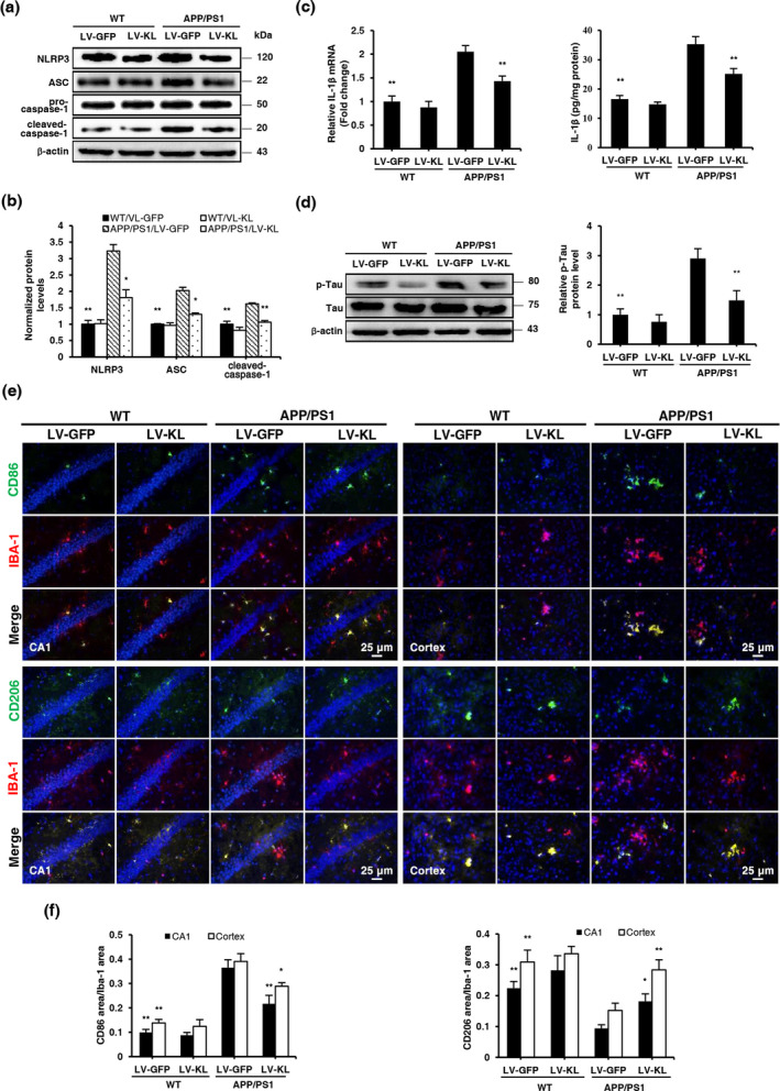
FIGURE 4.

Klotho overexpression promoted microglial transformation and alleviated Tau pathology in the brain in aged APP/PS1 mice. (a, b) Representative Western blotting and quantification of NLRP3, ASC, cleaved caspase‐1, and β‐actin in brain tissues. The amount of NLRP3, ASC, and cleaved caspase‐1 were normalized to β‐actin. (c) Analyses of IL‐1β mRNA levels and protein concentrations in brain homogenates by qRT‐PCR and ELISA, respectively. Relative mRNA levels of IL‐1β were normalized to GAPDH and are expressed as fold changes relative to the WT/LV‐GFP group. (d) Representative Western blotting and quantification of p‐Tau and Tau in brain tissues. The amount of p‐Tau was normalized to Tau. (e) Representative images of CD86 and CD206 (green) counterstained with Iba‐1 (red) and nuclear DNA staining of DAPI (blue) in the hippocampal CA1 area and cortex. (f) Quantitative image analysis of CD86/Iba‐1 and CD206/Iba‐1 expression based on positive fluorescence area in the hippocampal CA1 area and cerebral cortex. The data are expressed as each normalized value relative to the WT/LV‐GFP group. n = 6/group, except n = 4/group in (a, b, d). The data are expressed as mean ± SEM. The statistical analysis was performed using two‐way ANOVA followed by the Bonferroni‐Holm post hoc test. *p < 0.05, **p < 0.01, vs. APP/PS1/LV‐GFP group
