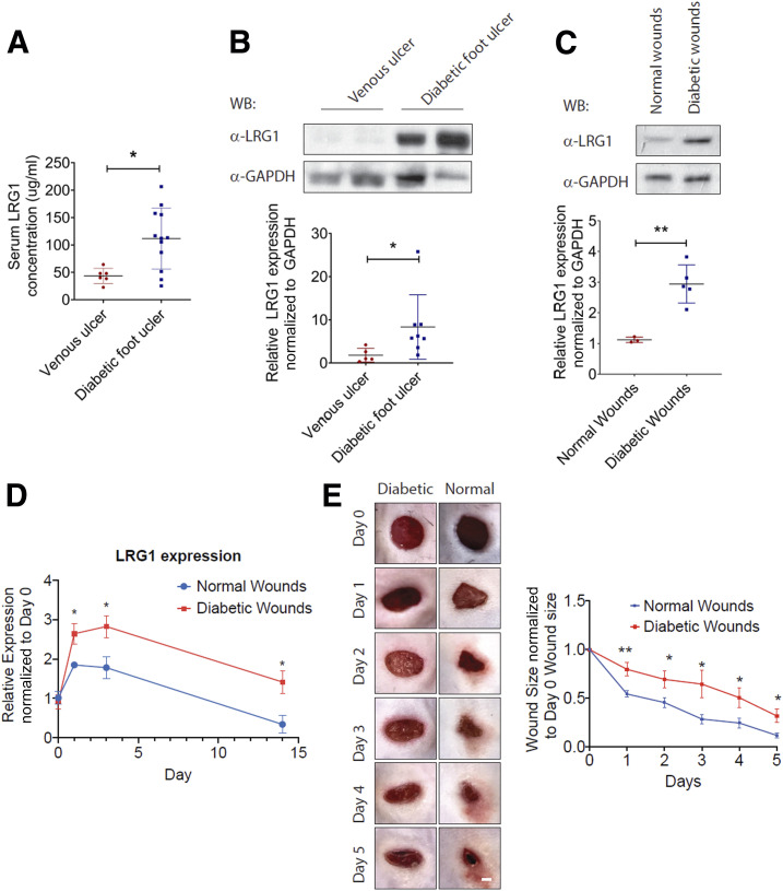
Figure 6.
Elevated LRG1 expression is observed in diabetic humans and mice. A: ELISA analysis of LRG1 in serum from venous ulcer patients and DFU patients. B: Representative Western blot (top) and densitometry analysis (bottom) of LRG1 and GAPDH in human patients with venous ulcer and DFU. C: Representative Western blot (top) and densitometry analysis (bottom) of LRG1 and GAPDH in normal and diabetic wounds of C57BL/6 mice. D: qRT-PCR analysis of normal and diabetic wounds of C57BL/6 mice. E: Representative images (left) and quantification (right) of wound size revealed a delayed wound closure in C57BL/6 mice with STZ-induced diabetes. Scale bar: 1 mm. All images are representative, and data are represented as mean (95% CI; P) of n ≥ 6 patients or mice per group. Significance was determined by unpaired, two-tailed Student t test. *P < 0.05, **P < 0.01. WB, Western blot.

