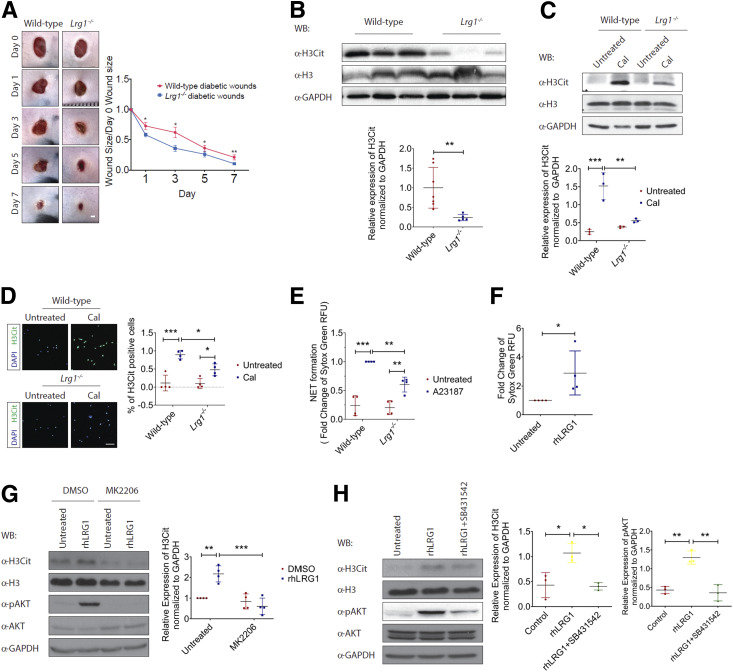
Figure 7.
LRG1 mediates NETosis. A: Representative images (left) and quantification (right) of wound size in wild-type and Lrg1−/− mice with STZ-induced diabetes. Scale bar: 1 mm. B: Representative Western blot (top) and densitometry analysis (bottom) of H3Cit, histone H3 (H3), and GAPDH in day 3 wounds from wild-type and Lrg1−/− mice with STZ-induced diabetes. C: Representative Western blot (top) and densitometry analysis (bottom) of H3Cit, H3, and GAPDH in calcium ionophore–treated wild-type and Lrg1−/− neutrophils. D: Representative immunofluorescence staining detecting H3Cit (green) and DAPI (blue) (left) and quantification of percentage of H3Cit+ cells (right) in calcium ionophore–treated wild-type and Lrg1−/− neutrophils. Scale bar: 80 μm. E: SYTOX Green assay on calcium ionophore–treated wild-type and Lrg1−/− neutrophils. F: SYTOX Green assay on calcium ionophore–treated dHL-60 cells. G: Representative Western blot (left) and densitometry analysis (right) of H3Cit, H3, AKT, phospho-AKT (pAKT), and GAPDH in rhLRG1- and/or MK2206-treated dHL-60 cells. H: Representative Western blot (left) and densitometry analysis (right) of H3Cit, H3, AKT, phospho-AKT, and GAPDH in rhLRG1 with or without SB431542-treated dHL-60 cells. All images are representative, and data are represented as mean (95% CI; P) of n ≥ 5 mice or n ≥ 3 independent experiments per group. Significance was determined by one- or two-way ANOVA followed by Tukey multiple comparisons test or unpaired, two-tailed Student t test. *P < 0.05, **P < 0.01, ***P < 0.001. CaI, calcium ionophore; WB, Western blot.

