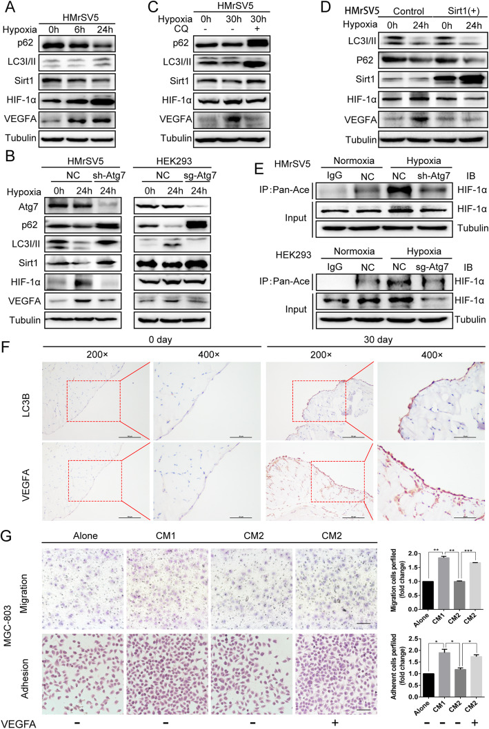
Fig. 6.
Hypoxia-induced autophagy mediated degradation of SIRT1 in PMCs promotes VEGFA secretion through acetylation of HIF-1α. a The levels of p62, LC3I/II, SIRT1, HIF-1α, and VEGFA were observed in HMrSV5 cells cultured in hypoxic conditions for the indicated time. b and c Western blot analysis of p62, LC3I/II, SIRT1, HIF-1α, and VEGFA under hypoxia and in response to treatment with chloroquine (CQ) or knockdown of ATG7 in HMrSV5 cells, or knockout of ATG7 in HEK293. d The levels of LC3I/II, p62, SIRT1, HIF-1α, and VEGFA were analyzed in HMrSV5 cells exposed to normoxia or hypoxia for 24 h with or without overexpression of SIRT1. e ATG7 was knocked out in HEK293 cells and knocked down in HMrSV5 cells, followed by exposure to normoxic or hypoxic conditions for 24 h. Immunoprecipitation was performed with a pan-acetyl antibody followed by immunoblot analysis using antibodies against HIF-1α. f Immunohistochemical analysis of LC3BI/II and VEGFA in benign mouse peritonea and GC metastatic peritonea. G. MGC-803 cells were subjected to normal media or conditioned media (CM1: conditioned media from hypoxic PMCs, CM2: conditioned media of hypoxic shRNA-Atg7 PMCs) and to CM2 with synchronous addition of exogenous VEGFA. Representative photographs of adherent and migratory cells are shown. Scale bars represent 100 μm. Bars represent SD of the mean. *P < 0.05. **P < 0.01. ***P < 0.001

