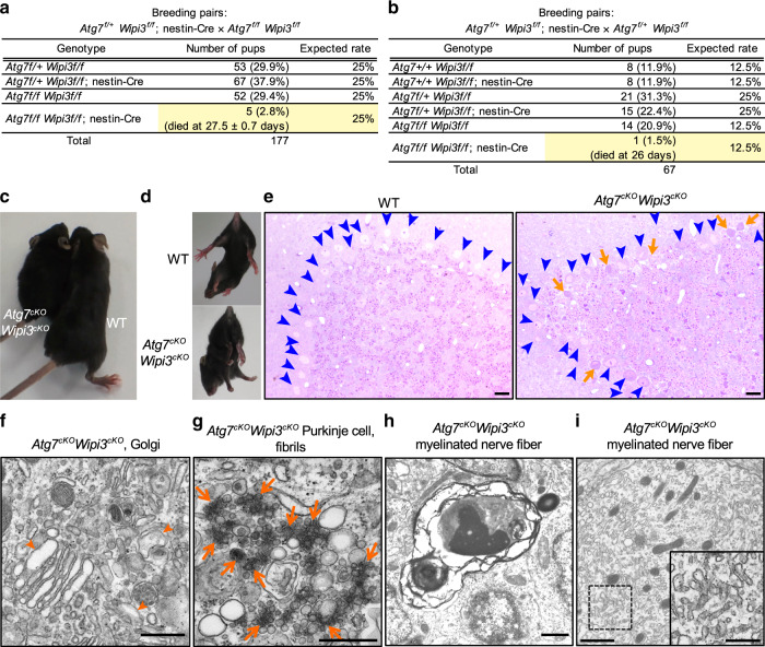
Fig. 10. Atg7cKO/Wipi3cKO mice showed a more severe phenotype than single-knockout mice.
a, b Tables showing the rate of viable neonates of the different genotypes. “Expected rate” indicates the Mendelian ratio. Yellow rows indicate the population of Atg7cKO/Wipi3cKO mice. c Gross appearance of the Atg7cKO/Wipi3cKO mice at 4 weeks of age. d The limb-clasping reflex is observed in Atg7cKO/Wipi3cKO mice. e Histological analyses of Atg7cKO/Wipi3cKO mice. Semi-thin sections of the cerebellum were stained with toluidine blue. Bars = 20 µm. Blue arrowheads and orange arrows indicate live and dead Purkinje cells, respectively. f–i EM analysis of Atg7cKO/Wipi3cKO mouse brains. f Swollen rod-shaped Golgi membranes were evident in the Purkinje cells from Atg7cKO/Wipi3cKO mice (arrowheads). The morphology was similar to that of Wipi3cKO mouse brains (Fig. 7i). g The accumulation of fibrils was observed in Purkinje cells from Atg7cKO/Wipi3cKO mice (arrows), as observed in Wipi3cKO Purkinje cells (Fig. 7k). h, i Myelinated nerve fibers of Atg7cKO/Wipi3cKO cerebella were filled with debris (h) and smooth ER (i). Bars = 0.5 µm in (f, g) and 1 µm in (h, i). Magnified images of the dashed squares are shown in the insets. Bar = 0.5 µm.

