
Figure 2.
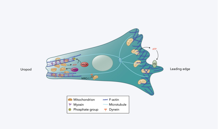
Mitochondrial redistribution in a migrating immune cell. At the leading edge, a small proportion of mitochondria with relatively high membrane potential accumulates. ATP derived from these mitochondria is released extracellularly and acts on the cell's purinergic P2 receptors (P2X4 and P2Y2 receptors in T cells and neutrophils, respectively), promoting the formation of actin protrusions. At the cell rear (uropod), there is localization of a larger fraction of mitochondria with lower membrane potential that are regulated by DRP1. These mitochondria are redistributed by coupling to dynein motor proteins on microtubules by MIRO‐1. They facilitate contraction of the uropod by providing ATP for myosin light chain kinase (MLCK) phosphorylation, and thus, MLC activation in T cells.