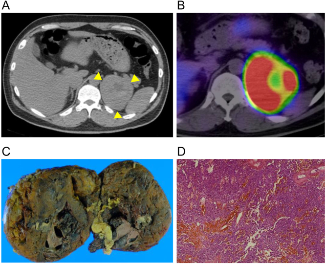
Figure 1.
Imaging findings. (A) Computed tomography with iodinated contrast media shows internal necrosis of the large adrenal mass (yellow arrows). (B) 123I-metaiodobenzylguanidine scintigraphy shows extensive accumulation in the adrenal tumor. The site of necrosis appears like a doughnut with no accumulation. (C) Macroscopic and (D) microscopic findings of the tumor.

 This work is licensed under a