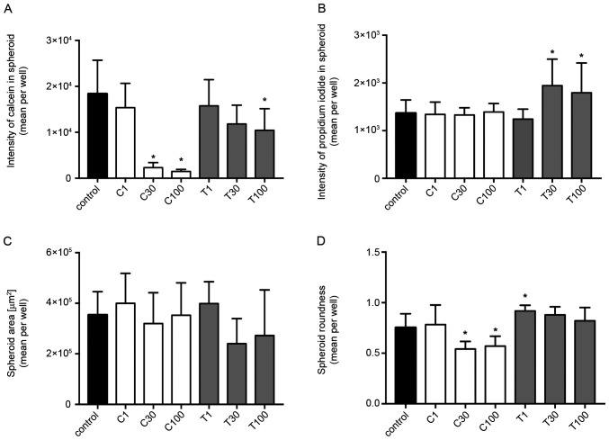
Figure 8.
Fluorescence intensities and morphological examinations of spheroids of 3D MDA-MB-231 cells treated with different concentrations of CEP or TET. Fluorescence intensity of (A) calcein and (B) propidium iodide in spheroids. (C) Spheroid area and (D) spheroid roundness of 3D MDA-MB-231 cells treated with CEP or TET. The intensity of calcein (cytoplasm) and propidium iodide (dead cells) fluorescence were detected after 15 min of incubation using an Operetta CLS (PerkinElmer, Inc.). The spheroid area and the roundness were calculated using the same instrument. The data are shown as the mean + SD of 4 independent experiments. A roundness value of 1 corresponds to complete roundness, and lower values indicate the deformation of the spheroid. *P<0.05 vs. control analyzed via one-way ANOVA and Dunnett's multiple comparisons test. C/CEP, cepharanthine; T/TET, tetrandrine.

