FIG 4.
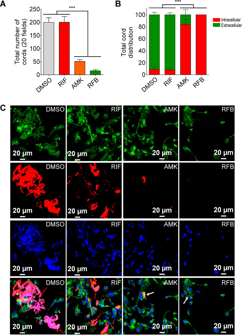
Activity of RFB on extracellular and intracellular cords. (A) Total number of cords displayed in 20 fields at 3 days after infection of macrophages with M. abscessus R variant. Data are mean values ± SD for three independent experiments performed in triplicate. Data were analyzed using one-tailed Mann-Whitney’s t test. (B) Percentage of cords formed either extracellularly or intracellularly. The two categories were counted at 3 days postinfection in the absence of antibiotics or in the presence of 50 μg/ml RIF, 50 μg/ml AMK, or 12.5 μg/ml RFB. Extracellular or intracellular cords are highlighted using the indicated color codes. Values are means ± SD for two independent experiments performed each time in triplicate. (C) Four immunofluorescent fields were taken at 3 days postinfection showing the cords formed extracellularly or within macrophages infected with M. abscessus R variant expressing Tdtomato (red). Macrophages were infected for 3 days in the presence of DMSO, RIF (50 μg/ml), AMK (50 μg/ml), or RFB (12.5 μg/ml). The macrophage surface was stained using anti-CD63 antibodies (green). The nuclei were stained with DAPI (blue). White arrows indicate intracellular cords, while red arrows indicate extracellular cords. Scale bars represent 20 μm. Results represent the average of a total of 120 fields per condition. ****, P ≤ 0.0001.

