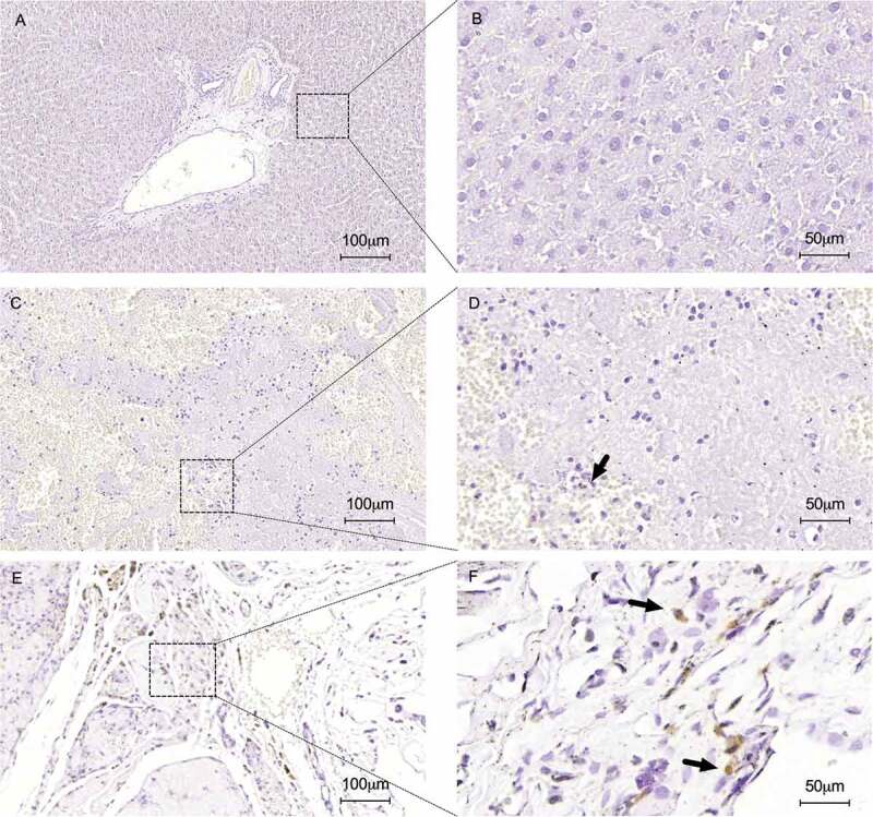
Figure 11.

Immunohistochemical staining for apoptotic nuclei detection using the TUNEL method. No apoptotic nuclei were detected in normal liver sections represented in low (a) and high magnification (b). TUNEL analysis confirmed apoptotic cells into heterotopically (c and d) and orthotopically (e and f) transplanted ALS (black arrows). Normal liver samples (a and b) were used such as control. Scale bars: 100 µm (A, C and E), 50 µm (B, D and F), respectively.
