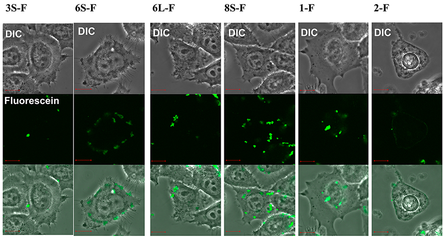
Figure 8.

Imaging experiments for cell permeability of the stapled and linear peptides labeled with fluorescein to HeLa cells. Each panel is divided into three sections as follows: upper, differential interference contrast (DIC) image; middle, fluorescein emission; lower, merged image. The peptide concentration is 5 μM. After addition of peptides, cells were incubated at 37 °C for 30 min under 5% CO2 atmosphere. After washing three times with HBSS buffer, the fluorescent imaging was conducted by Fluoview FV10i confocal microscopy systems (Olympus). Orange bars in the panels represent 10 μm. The fluorescein-labeled peptides contain fluorescein–GABA instead of acetyl in the N-terminus.
