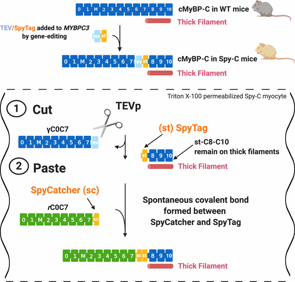
Figure 1. A “cut and paste” method for replacing N’-terminal domains of cMyBP-C in situ.
Top, cMyBP-C consists of 11 folded domains numbered C0 to C10 starting at the N’-terminus of the molecule plus an additional “M”-domain unique to MyBP-C proteins between C1 and C2. Gene-edited Spy-C mice express a modified cMyBP-C with a TEV protease (TEVp) recognition site (light blue rectangle) and a SpyTag (orange rectangle) inserted between domains C7 and C8. (1) CUT: TEVp treatment of detergent-permeabilized homozygous (HO) Spy-C myocytes releases genetically encoded (γ)C0-C7. (2) PASTE: New recombinant (r)C0-C7-sc domains (green) (encoding any desired modification such as point mutations, deletions, fluorescent probes) are added to the bath where they are covalently attached to st-C8C10 on the thick filament via a spontaneous bond formed between SpyCatcher and SpyTag. Figure created with BioRender.com.

