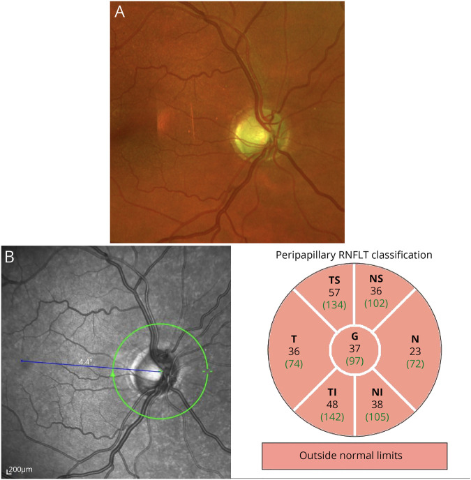
Figure. Ophthalmologic findings.
(A) Ultrawide field photography performed on the Optos system (Dunfermline, UK) showed a sharp and pale optic disc with a cup-disc-ratio of 0.4 and a subtle peripapillary atrophy. (B) The examination of the retinal nerve fiber layer on optical coherence tomography (Spectralis OCT, Heidelberg Engineering, Heidelberg, Germany) showed a pronounced loss of nerve fibers in all quadrants.

