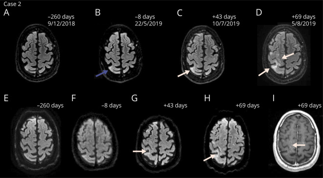
Figure 3. Magnetic resonance images demonstrating the disease course of PML-IRIS of case 2.
Axial FLAIR (A–D), diffusion-weighted (E–H) and contrast-enhanced T1-weighted (I) magnetic resonance images of PML with IRIS of case 2. In July 2019, new focal areas with elevated signal on the diffusion weighted imaging setting in the right pre- and postcentral gyrus and gyrus supramarginalis were detected (C and G). Follow-up scans showed multiple foci of elevated signal suspect for PML-IRIS (D, H, I). The amount of days indicates the time from the first infusion with ocrelizumab. Natalizumab was discontinued in April 2019 and PML was diagnosed after 97 days. Blue arrow indicates subtle signs suggestive of PML in retrospect before diagnosis (B). White arrows indicate the lesions suggestive of PML-IRIS. FLAIR = fluid-attenuated inversion recovery; IRIS = immune reconstitution inflammatory syndrome; PML = progressive multifocal leukoencephalopathy

