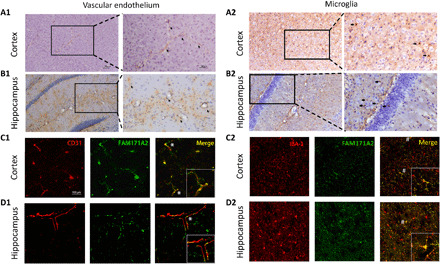
Fig. 5. FAM171A2 high expression on cerebral vascular endothelium and microglia.

(A and B) The IHC staining of FAM171A2 on mouse cortex and hippocampi. The DAB staining along and around the cerebral vascular (A1 and B1) was marked by tilted arrows. The DAB staining on the cells in a similar form of microglia (A2 and B2) was marked by horizontal arrows. (C and D) The IF staining of FAM171A2 with the CD31 and IBA1 antibodies on mouse cortex and hippocampus. Their colocalization was marked by “*” and “#.” n = 5 mice in these experiments.
