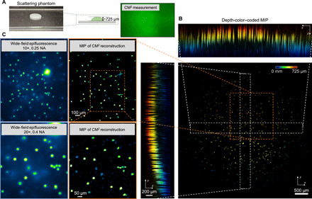
Fig. 6. Imaging of a scattering sample with a curved surface.

(A) Illustration of the scattering sample (ls ~ 264 μm) with a curved surface and CM2 raw measurement. Photo credit: Yujia Xue, Boston University. (B) The depth-coded MIPs of the CM2 reconstruction recovers particles in the superficial layer of the curved surface. (C) The comparison between the CM2 reconstruction and the wide-field fluorescence measurements (10×, 0.25 NA and 20×, 0.4 NA) verifies that the CM2 correctly reconstructs the emitters in the superficial layer.
