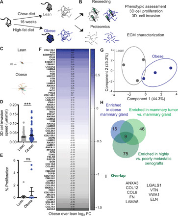
Fig. 2. ECM from the mammary gland of obese mice increases cell invasion and overlaps with breast tumor ECM composition.

(A) Diet-induced obesity model in female C57BL/6 mice. (B) dECM of lean and obese mammary glands by decellularization was used for reseeding and proteomics. Representative tracks (C) and quantification (D) of cell migration speed of 231-GFP cells seeded on dECM from lean and obese mammary gland. Each point represents the average speed of a cell over 16 hours. (E) Percentage of proliferating 231-GFP cells on ECM scaffolds from lean and obese mammary gland. Data show mean ± SEM, obtained from at least three dECM scaffolds from three mice. Significance was determined by unpaired two-tailed Mann-Whitney t test. ***P ≤ 0.005. (F) Average log2 fold change (FC) differences of 68 ECM proteins identified by proteomics of the ECM fraction and principal components analysis (G). (H) ECM proteins found enriched in obese mammary gland were overlapped with proteins enriched in 4T1 orthotopic syngeneic mammary (9) tumor and in MDA-MB-231-LM2 highly metastatic (10) xenografts in a Venn diagram. (I) List of nine matrisome proteins present in obese mammary gland ECM, 4T1 tumor ECM, and metastatic LM2 tumors.
