. 2020 Oct 5;9:e51898. doi: 10.7554/eLife.51898

Figure 2. Identity of the mRNA proline codon regulates 30S head domain swivel and tilting.

Overview of 70S ribosome-tRNAPro complex structures: (A) tRNAPro m1G37 on a cognate CCG codon adopts a P/P orientation (located on the P site on the 30S and 50S subunits); (B) tRNAPro lacking the m1G37 modification (G37 Δm1) on a cognate CCG codon also adopts a P/P orientation; (C) tRNAPro on a +1 slippery CCC-U codon adopts an e*/E orientation (e* denotes the location between the E and P sites on the 30S while "E" is the E site of the 50S); and (D) tRNAPro lacking the m1G37 (G37 Δm1) on a +1 slippery CCC-U codon adopts an e*/E orientation. In this complex (panel D), the 30S head domain and anticodon-codon interaction are disordered. In panels A-D, the 16S rRNA of the 30S head domain is removed for clarity. Zoomed-in view of 2Fo-Fc density of (E) the codon–anticodon for tRNAPro on a cognate CCG codon, (F) tRNAPro G37 (Δm1) on a cognate CCG codon in the P site, and (G) tRNAPro G37 (Δm1) on a near-cognate CCC-U codon in position between the E and the P sites (e*). All 2Fo-Fc electron density maps shown in panels E-G are contoured at 1.0σ.

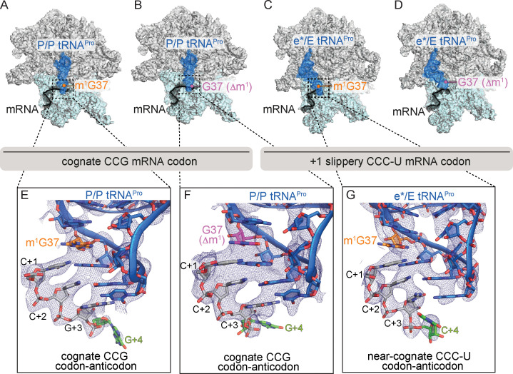
Figure 2.

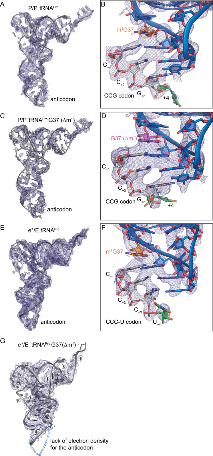
Figure 2—figure supplement 1. mRNA and full-length tRNAProelectron density.

Figure 2—figure supplement 1.

(A) 2Fo-Fc electron density for full-length tRNAPro on a cognate codon in the P site. (B) Zoomed-in view of codon–anticodon 2Fo-Fc density for full-length tRNAPro on a cognate CCG codon. (C) 2Fo-Fc density for full-length tRNAPro G37 (Δm1) on a cognate codon in the P site. (D) Zoomed-in view of codon–anticodon 2Fo-Fc density for full-length tRNAPro G37 (Δm1) on a cognate CCG codon. (E) 2Fo-Fc density for full-length tRNAPro on a near-cognate codon in the e* site. (F) Zoomed-in view of codon–anticodon 2Fo-Fc density for tRNAPro on a near-cognate CCC-U codon. (G) 2Fo-Fc density for 16S rRNA and full-length tRNAPro G37 (Δm1) on a near-cognate CCC-U codon in the e* site. The portion of the ASL that lacks density is represented as a hollow blue dashed line. 2Fo-Fc density is is contoured at 1σ for all structures. In the zoomed-in views of panels B, D, and F, the +four position on the mRNA is highlighted in green to indicate the structures are in the 0 frame.
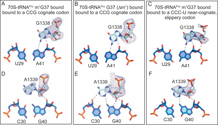
Figure 2—figure supplement 2. Full-length tRNAPro interactions with 16S nucleotides G1338 and A1339.

Figure 2—figure supplement 2.

Comparison of the A-minor motif interactions of G1338 with tRNAPro U29-A41 base-pairs (A) and A1339 with tRNAPro C30-G40 base-pairs in (D) 70S-tRNAPro bound to a cognate CCG codon. (B,E) 70S-tRNAPro G37 (Δm1) bound to a cognate CCG codon. (C,F) 70S-tRNAPro bound to a near-cognate CCC-U codon. 2Fo-Fc density for G1338 and A1339 is contoured at 1σ.
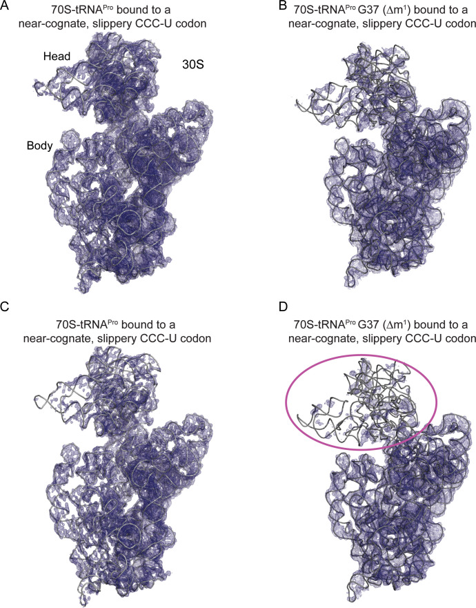
Figure 2—figure supplement 3. tRNAPro G37 (Δm1) coupled with a slippery codon capable of +1 frameshifting induces disorder in the 30S head domain.

Figure 2—figure supplement 3.

(A) 2Fo-Fc density at 1.0σ of 16S rRNA in the 70S-tRNAPro bound to a cognate CCG codon. (B) 2Fo-Fc density at 1.0σ of 16S rRNA in the 70S-tRNAPro G37 (Δm1) bound to a near-cognate CCC-U codon. (C,D) The same ribosome complexes as in panels A and B but with 2Fo-Fc electron density increased to 1.5σ.