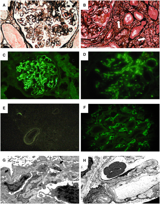
FIGURE 2.
In a patient with recurrent IgAN, the light microscopy picture (A) shows a glomerulus with global membranoproliferative pattern of injury with several aspects of glomerular basement membrane double contours as well mesangial and capillary wall eosinophilic deposits (black arrow) (Jones silver stain). In this patient, immunofluorescence microscopy revealed mesangial and glomerular capillary wall deposition of IgA (C) with negative C4d in the peritubular capillaries (E). Electron microscopy in this case showed subendothelial electron dense deposits (black arrows) with newly formed lamina densa (i.e. double contours) and cellular interposition (white arrow) (G) (US, urinary space; CL, capillary lumen) [transmission electron microscopy (TEM), ×8900]. In a patient with recurrent IgAN and concurrent antibody-mediated rejection, a light microscopy image (B) shows a portion of a glomerulus (on the left) with segmental obliteration of CLs due to endothelial cells swelling and mononuclear inflammatory cells (i.e. glomerulitis) with inflammatory cells also in peritubular capillaries (white arrows) (Jones silver stain). In this patient, immunofluorescence microscopy revealed mesangial deposition of IgA (D) with diffuse strongly positive C4d in the peritubular capillaries (F). Electron microscopy of this case showed widening of the subendothelial spaces in the absence of subendothelial deposits (TEM, ×8900).

