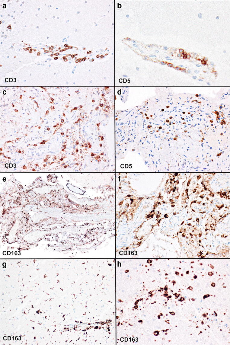
Fig. 7.
a Perivascular inflammatory infiltrates composed partly of CD3-immunopositive lymphocytes in the cerebral cortex. b Lymphocytes are also immunopositive for CD5. c Leptomeningeal infiltrates of CD3-immunopositive lymphocytes. d Lymphocytes are also immunopositive for CD5. e, f Leptomeningeal infiltrate of mostly perivascular CD163-immunopositive histiocytes. g, h Perivascular infiltrate of CD163-immunopositive histiocytes and numerous microglia in the cerebral cortex. (a and c CD3 immunostains, b and d CD5 immunostains, e–h CD163 immunostains; original magnification a and h × 400; b × 800; c, d, and f × 200; e × 40; g × 100)

