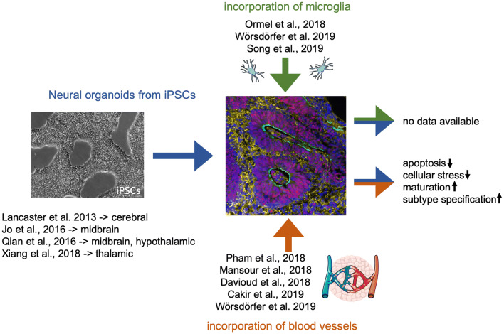
Fig. 2.
Incorporation of microglia and blood vessels into neural organoids. This fig. shows recent publications describing (1) the generation of brain region specific neural organoids (Jo et al. 2016; Qian et al. 2016; Xiang et al. 2019), (2) the incorporation of microglia into neural organoids and (3) the incorporation of a vascular system into neural organoids as well as the so far demonstrated advantages of increasing organoid complexity. The phase contrast image (left side) shows undifferentiated iPSCs in 2D culture. The immunofluorescence image (right side) shows an immature neural organoid generated from hiPSCs stained for Sox1 (neural stem cells—> red), MAP2 (neurons—> yellow), and N-Cadherin (green). Graphical elements within this schematic were taken from the image bank from Servier Medical Art licensed under a Creative Commons Attribution 3.0 Unported License (https://creativecommons.org/licenses/by/3.0)

