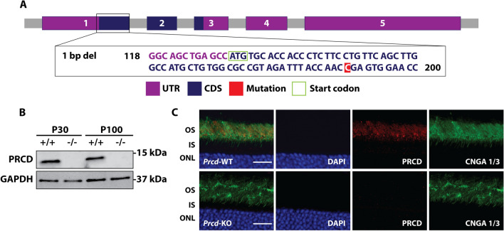
Figure 1.
Generation and validation of Prcd-KO animal model. (A) Scheme demonstrating the generation of the Prcd-KO animal model using CRISPR/Cas9 technology. In this study, we used the 1738 founder line, which has a single base pair deletion in exon 1 of Prcd. UTR = untranslated region, CDS = coding sequence. (B) Immunoblot analysis shows loss of PRCD protein from Prcd-KO retinal lysate at postnatal (P) 30 and P100, whereas wild-type (WT) retinal lysates demonstrate PRCD immunoreactivity (n = 4). Please note that immunoblot data is cropped; full-length, raw data is available in Supplementary Fig. S2. (C) Immunofluorescent staining of P30 retinal cross-sections from WT and Prcd-KO mice, probing with antibodies against PRCD (red) and the OS marker cyclic nucleotide gated channel alpha-1 and alpha-3 (CNGA 1/3; green) to demonstrate proper localization of PRCD to the OS in WT retina and loss of PRCD from Prcd-KO retina (n = 3). Scale bar = 20 μm. All experiments were conducted with littermate controls.

