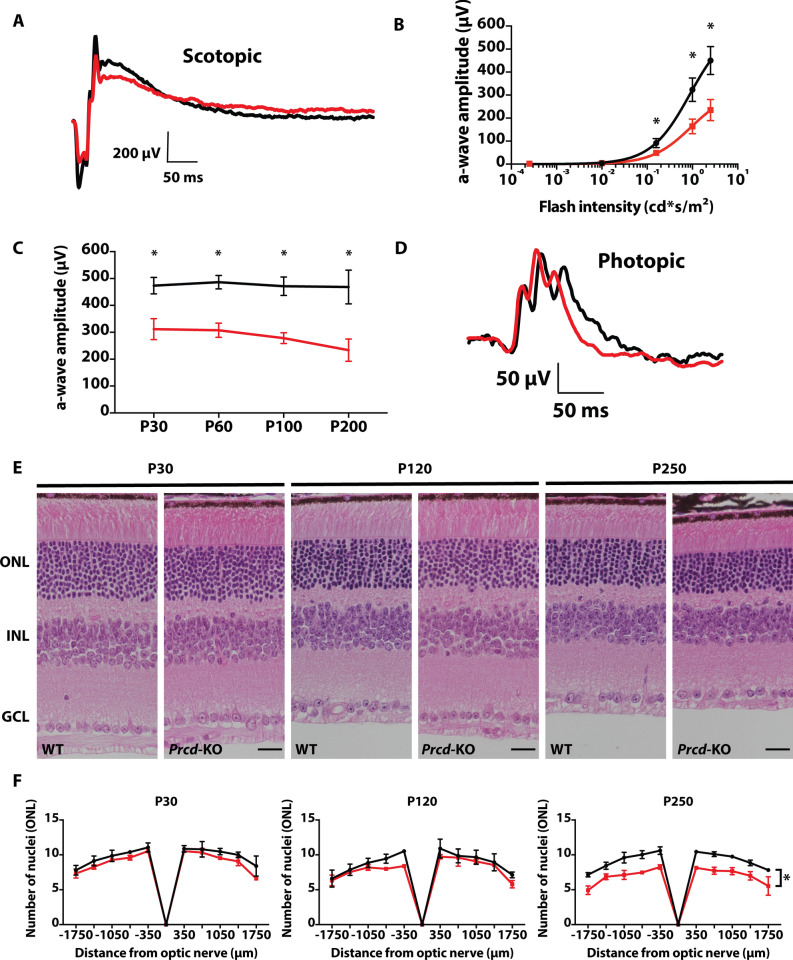
Figure 2.
Reduced rod photoreceptor function and slow retinal degeneration in animals lacking Prcd. (A) Representative waveform from scotopic ERGs of WT (black) and Prcd-KO (red) animals at P30 (0.995 cd*s/m2). (B) Significant loss of rod photoreceptor function at higher light intensities (0.158, 0.995, and 2.5 cd*s/m2) compared with low light conditions (0.00025 and 0.001 cd*s/m2) (n = 4, data from B and C stats are unpaired two-tailed t-test; higher light intensities were statistically significant, *p < 0.01; Low light intensities were not significant). (C) Maximum a-wave amplitude of Prcd-KO animals at different ages compared to WT controls from P30 to P200. (D) Representative waveform from photopic (cone) ERGs of Prcd-KO and WT animals at P200 (7.9 cd*s/m2) (n = 4). (E) Prcd-KO and WT littermate control cross-sections stained with hematoxylin and eosin (H&E), imaged by light microscopy, at P30, P120, and P250. Scale bar = 20 μm. (F) Quantification of number of photoreceptor nuclei in the outer nuclear layer (ONL) from both Prcd-KO (red) and WT littermate controls (black), every 350 μm from the optic nerve, at P30, P120, and P250. Data are represented as mean (n = 3, unpaired two-tailed t-test; *p < 0.01).

