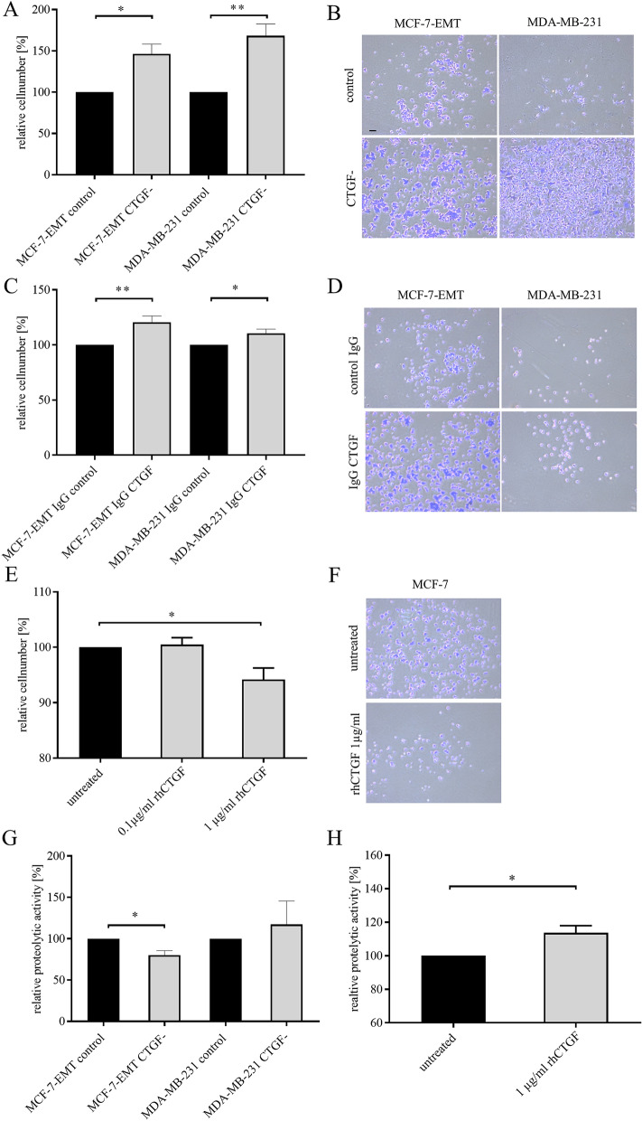
Figure 4.
CTGF alters cell-ECM adhesion and proteolytic activity of breast cancer cells. (A) Adhesion analysis of transiently transfected mesenchymal transformed and triple-negative breast cancer cells. Adhesive cells where counter-stained with crystal violet and absorption was measured at 570 nm. Data represent mean ± SEM. MCF-7-EMT n = 3, MDA-MB-231 n = 3 using unpaired, two-tailed t-test analysis to respective control. *P < 0.05; **P < 0.01 (B) Representative images corresponding to A. (C) Extracellular CTGF was reduced using a blocking-antibody against CTGF and cell-ECM adhesion was assessed. Data represent mean ± SEM. MCF-7-EMT n = 6, MDA-MB-231 n = 3 using unpaired, two-tailed t-test analysis to respective control (IgG control). *P < 0.05; **P < 0.01 (D) Representative images corresponding to C. (E) MCF-7 cells where treated with recombinant human CTGF (rhCTGF) in different concentrations prior to assessing of cell-ECM adhesion. Data represent mean ± SEM. n = 3 using one-way ANOVA with F = 6.244 and a Dunnett ‘s multiple comparison test with no matching or pairing between groups. *P < 0.05 (F) Representative images corresponding to E. (G) Following transient transfection mesenchymal transformed and triple negative breast cancer cells were seeded on FITC-conjugated gelatin (0.2%). Degradation of gelatin /proteolytic activity results in an increase of fluorescence. Data represent mean ± SEM. MCF-7-EMT n = 3, MDA-MB-231 n = 3 using unpaired, two-tailed t-test analysis to respective control. *P < 0.05 (H) Assessment of proteolytic activity of MCF-7 breast cancer cells after treatment with rhCTGF. Data represent mean ± SEM. M n = 3 using unpaired, two-tailed t-test analysis to respective control (untreated).*P < 0.05. Scale bar gauges 200 µm.

