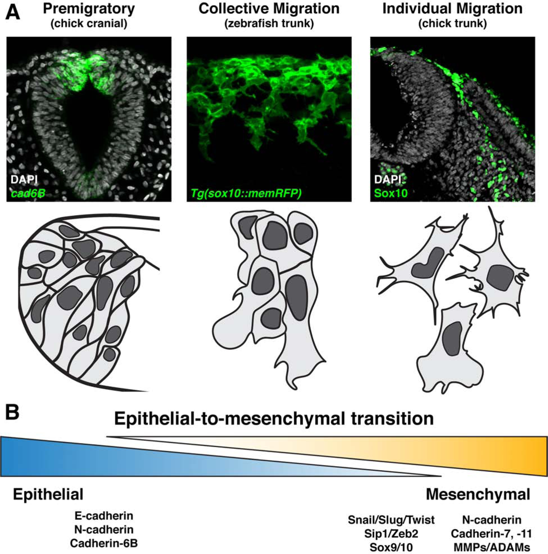
Figure 2. Neural crest epithelial-to-mesenchymal transition produces migratory cells of varying degrees of mesenchymalization.

(a) Representative images (top panels) and schematics (bottom panels) displaying neural crest cells at varying degrees of epithelial-to-mesenchymal transition. Premigratory neural crest cells reside in the neuroepithelium exhibit highly epithelial characteristics including tight cell adhesions, as demonstrated by cadherin6B expression in chick cranial neural crest (left). Collective migration of neural crest cells occurs when there is a partial downregulation of epithelial, and upregulation of mesenchymal, characteristics. The resulting migratory cells traverse their environment as tight clusters or in chains with extensive cell-cell contacts, as illustrated by migrating zebrafish trunk neural crest (center). In contrast, individually migrating neural crest cells are more completely mesenchymalized and move through their migratory paths as free cells with more infrequent and transient interactions with one another, as is evident in the trunk of chick embryos (right). (b) Epithelial-to-mesenchymal transition is best considered as a spectrum of cell states ranging from epithelial to mesenchymal, rather than a binary switch between the two. Listed are a selection of common gene signatures expressed in more epithelial (left) and more mesenchymal (right) neural crest cells.
