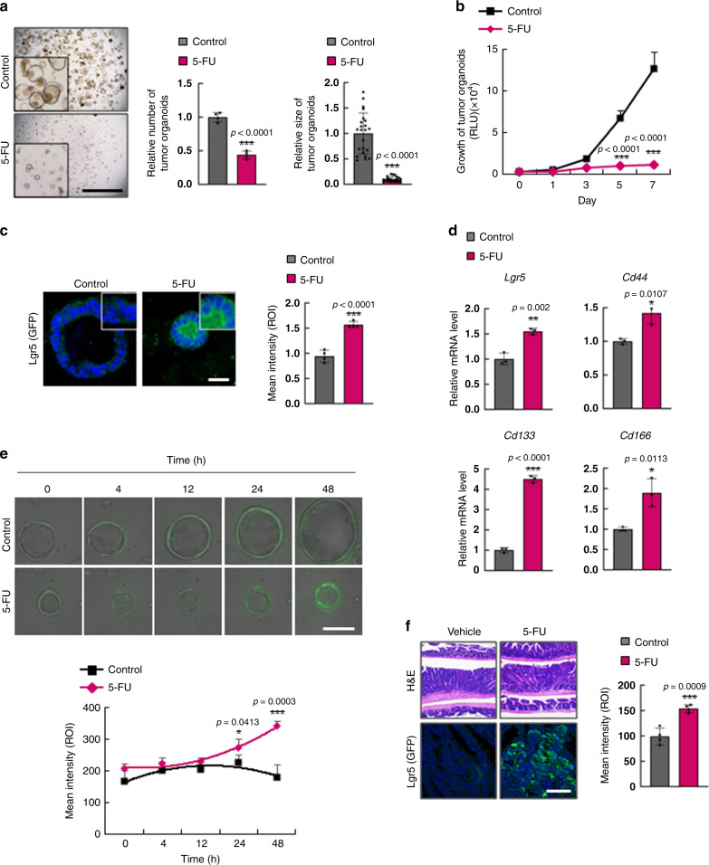
Fig. 1. 5-FU treatment induces CSC enrichment in murine CRC.
a–f Analyses of tumor organoids derived from intestinal tumors of ApcMin/+/Lgr5EGFP mice after treatment with 5-FU (1.5 μg/ml, 48 h). a Bright-field images of tumor organoids treated with 5-FU. Relative numbers (n = 4 biologically independent samples per group) and sizes (n = 23 biologically independent organoids per group) of tumor organoids were quantified as fold-change compared to control. Scale bar = 5 mm. b Growth of tumor organoids after treatment with 5-FU for indicated days. (n = 5 biologically independent samples per group) c Confocal images of Lgr5 (GFP) and the mean intensity of GFP after treatment with 5-FU. Scale bar = 50 μm. (n = 5 biologically independent samples per group.) d Relative mRNA levels of Lgr5, Cd44, Cd133, and Cd166 after 5-FU treatment. Data shown as fold-change compared to control. (n = 3 biologically independent samples per group.) e Time-course analyses of Lgr5 (GFP) expression after 5-FU treatment. Scale bar = 100 μm. (n = 4 biologically independent samples per group.) f Hematoxylin and eosin and immunofluorescence staining of mouse intestinal sections after treatment with vehicle or 5-FU (25 kg/ml, 3 weeks) using antibodies for the indicated proteins. The mean intensities were measured by Zen software 3.1. Scale bar = 50 μm. (n = 4 biologically independent samples per group) Data are mean ± s.d., two-sided Student’s t-test, *p < 0.05, **p < 0.01, ***p < 0.001. Source data are provided as a Source data file.

