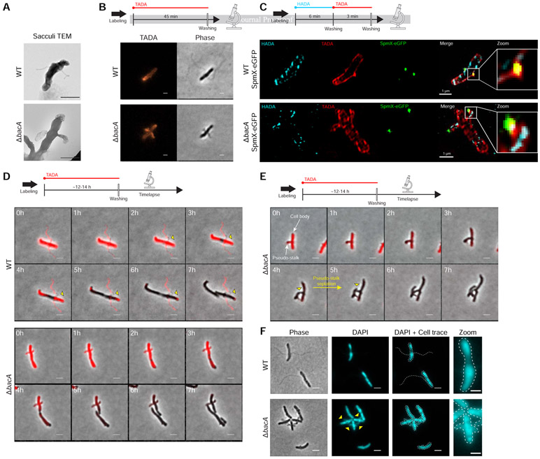
Figure 6. Peptidoglycan (PG) Composition & Remodeling and Septation & DNA Content in WT Stalks and ΔbacA Pseudostalks.
(A) Transmission electron microscopy (TEM) of prepared PG sacculi for strains YB642 (WT) and YB8597 (ΔbacA). Cells were grown in rich medium (PYE) to saturation and sub-cultured into phosphate limited (HIGG) medium at 26°C for 72h before preparing sacculi. Sacculi were prepared by boiling in SDS for 30 minutes followed by washing ≥6X with dH2O. Scale bars = 1 μm. (B) Medium pulse FDAA (TADA) labeling showing active PG remodeling in strains YB642 (WT) and YB8597 (ΔbacA). Cells were grown in rich medium (PYE) to saturation and sub-cultured into phosphate limited (HIGG) medium at 26°C for 72h ) before labeling. Cells were washed with 2X PYE, labeled with 500 μM TADA for 45 minutes, washed 2X with PYE, and imaged with phase and fluorescence microscopy. Representative images are shown. Scale bars = 2 μm. (C) Virtual time lapse: Short pulse, sequential, dual FDAA (TADA and HADA) labeling showing active PG remodeling in strains YB5692 (spmX::spmX-eGFP) and YB7561 (spmX::spmX-eGFP ΔbacA). Cells were grown in rich medium (PYE) to saturation and sub-cultured into phosphate limited (HIGG) medium at 26°C for 72h before labeling. Cells were washed 2X with PYE, labeled with 500 μM HADA for 6 minutes, washed 2X with PYE, labeled with 500 μM TADA for 3 minutes, washed 2X with PYE, and imaged via 3D-SIM (Structured Illumination Microscopy). Representative images are shown. Scale bars = 1 μm. (D) Pulse-chase time-lapse of FDAA (TADA) labeling for strains YB642 (WT) and YB8597 (ΔbacA) showing loss of labeling as peptidoglycan is actively remodeled. Yellow triangles with black outline indicate loss of TADA signal in YB642 (WT) as stalk is extended from the base. Cells were grown in rich medium (PYE) to saturation and sub-cultured into phosphate limited (HIGG) medium at 26°C for 60h before labeling. To label whole cells, 250 μM TADA was added and cells were allowed to grow an additional ~12-14h (overnight). Cells were then washed 2X with PYE to remove TADA and imaged via time-lapse with phase and fluorescence microscopy. Scale bars = 2 μm. (E) Pulse-chase time-lapse of FDAA (TADA) labeling for strains YB8597 (ΔbacA) showing elongation and septation of a pseudo-stalk. Yellow triangles with black outline indicate the septation site. Cells were grown as described in Figure 6D. Scale bars = 2 μm. (F) DAPI staining for DNA in strains YB642 (WT) and YB8597 (ΔbacA) showing DNA is present in the pseudo stalk (yellow triangles). Representative images are shown. Scale bars = 2 μm. See also Video S7.

