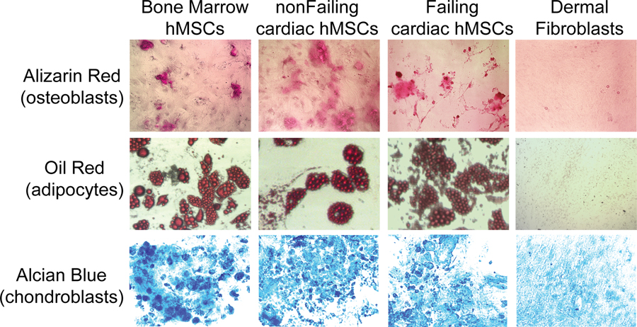
Figure 2:
Examples of osteoblasts (Alizarin Red, top), adipocytes (Oil Red, middle), and chondroblasts (Alcian Blue, bottom) differentiated from plastic adherent cells isolated from the bone marrow (positive control), a device lead extracted from a patient with a normal EF (non-failing hMSC, middle left) and a failing heart (Failing hMSC, middle right), and human dermal fibroblasts (negative control). After 3 weeks, a similar differentiation potential was observed in all groups, except dermal fibroblasts.

