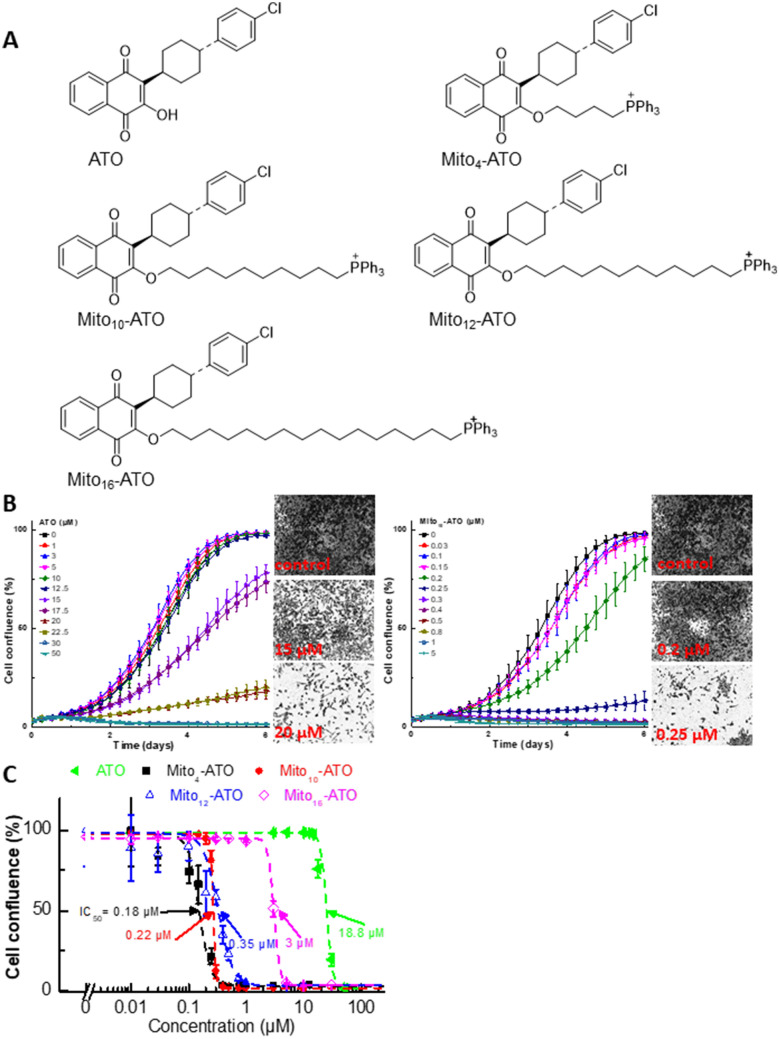
Figure 1.
Effects of ATO, Mito-ATO analogs, and related analogs on proliferation of MiaPaCa-2 cells. (A) Chemical structures of ATO and Mito-ATO analogs. (B) Effect of ATO on the proliferation of human pancreatic cancer cells was compared with that of Mito10-ATO in the IncuCyte Live-Cell Imager. MiaPaCa-2 cells were treated with ATO and Mito10-ATO. Cell proliferation was monitored in real-time with the continuous presence of indicated treatments until the end of each experiment. Created using NIH public domain image processing program, ImageJ38. (C) Cell confluence (as control groups reach 98% confluency) is plotted against concentrations of ATO and Mito-ATO analogs. Dashed lines represent the fitting curves used to determine their IC50 values as indicated.

