
Figure 4: Effect of color normalization on classifier performance.
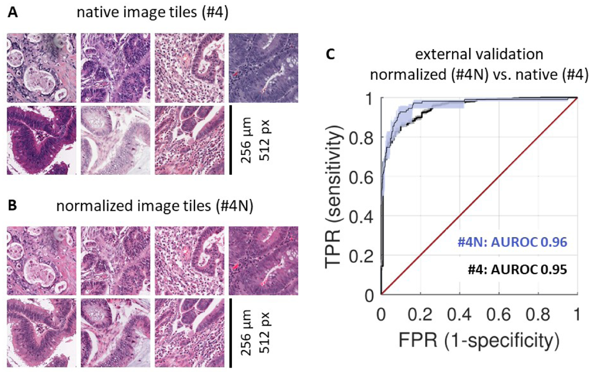
(A) A representative set of tiles from the MSIDETECT study. (B) The same tiles after color normalization. (C) Classifier performance on an external test set (YCR-BCIP-RESECT, n=771 patients) improves after color-normalizing training and test sets. Experiment #4N is with color normalization, experiment #4 is without color normalization. AUROC: area under the receiver operating curve, TPR: true positive rate, FPR: false positive rate.