
Figure 6.
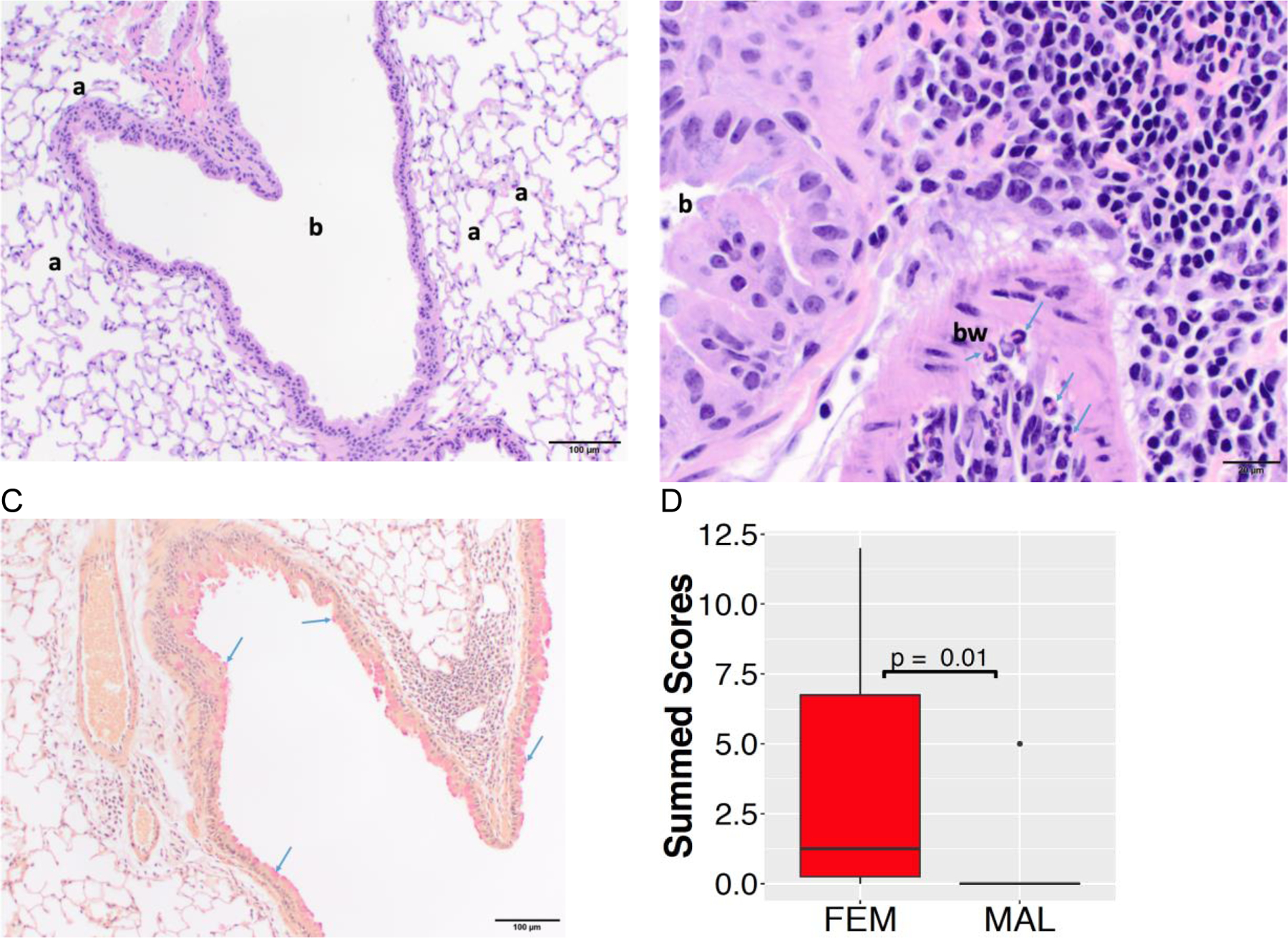
Lung histopathology in female and male offspring at PND 290. (A) Low magnification photomicrograph depicting a bronchiole (b) and surrounding alveoli (a) from a male mouse. The section is histologically normal. Hematoxylin and eosin staining, 100x original magnification, scale bar = 100µm. (B) High magnification photomicrograph of inflammatory infiltrate (right and top right sections of the image) around a bronchiole (b) and within the bronchiolar wall (bw) from a female mouse. Within the bronchiolar wall, there are numerous eosinophils (arrows). Hematoxylin and eosin staining, 400x original magnification, scale bar = 20µm. (C) Low magnification photomicrograph of mild goblet cell hyperplasia (bright red globules, arrows) in the bronchiolar epithelial lining from a female mouse. Mucicarmine staining, 100x original magnification, scale bar = 100µm. (D) The summed pathological score was higher in female offspring (FEM) than male offspring (MAL).