
Fig. 3.
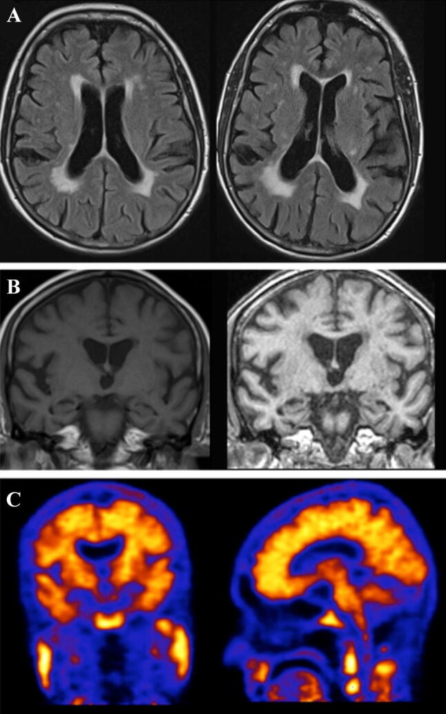
Case 3. Axial FLAIR images through the brain (a) demonstrating white matter changes over a 3-year interval. Coronal T1- and T2-weighted images through the brain (b) showing progressive atrophy of the brain over the 3-year interval. The most recent scan (right) shows bilateral medial temporal lobe atrophy, more so on the right. Amyloid PET imaging (c) was positive with loss of grey–white differentiation consistent with widespread amyloid deposition