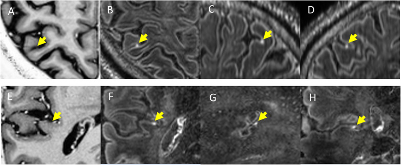
Fig. 2.
Examples of “nodular” type enhancing foci in multiple sclerosis. In the horizontal rows are displayed two different patients with multiple sclerosis. T1-weighted magnetisation-prepared two rapid gradient echo images (A, E), along with axial (D, H), sagittal (C, G), and coronal (B, F) slices from contrast-enhanced magnetisation-prepared fluid-attenuated inversion recovery images. The yellow arrows indicate the location of a nodular focus of leptomeningeal contrast enhancement in all three planes and its expected location on magnetisation-prepared two rapid gradient echo images. Published with permission from J Neurol [53]

