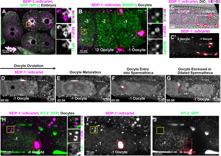
Fig. 4.
SEIP-1 localization in C. elegans oocytes and embryos. (A) SEIP-1::mScarlet observed in embryos. Right inserts represent the magnified area of the yellow square, showing that SEIP-1::mScarlet is adjacent to the ER labeled by GFP::SP12. (B) In the oocytes, SEIP-1::mScarlet is either adjacent to or surrounds the LDs stained by BODIPY. Right inserts represent the magnified area of the yellow square, showing that SEIP-1::mScarlet localizes to the surface of the BODIPY stained LDs. −1, −2 and −3 indicate oocyte from the most proximal to the distal to the spermathecal. (B-C′) SEIP-1::mScarlet localizes to the pseudocoelomic space (red squares in panel B; red circles in panels C,C′) surrounding the ovulating oocytes (-1 oocyte). (D-G) Representative images of SEIP-1::mScarlet localization during ovulation and fertilization. A strong SEIP-1::mScarlet signal was observed in the fifth sheath cell (red arrows) that surrounds the −1 oocyte (D) until the oocyte is ovulated and enters the spermatheca (E-G). (H-J) A vitellogenin reporter (VIT-2::GFP) transgene was used to monitor yolk lipoprotein localization, showing no colocalization with SEIP-1::mScarlet either in the LDs (yellow squares) or the pseudocoelomic space (red squares). Amplified images of the yellow square are shown to the right of H.

