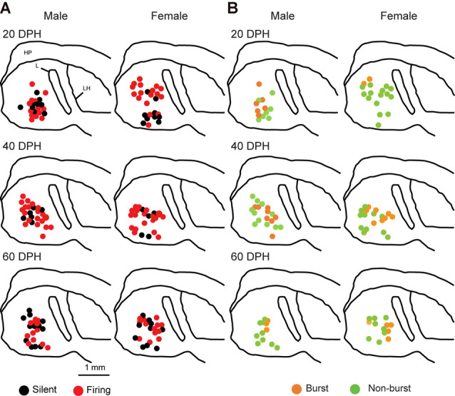
Figure 3.

Types of neurons with an SFR and burst-type were not correlated with a location within the NCM (X2 test). Anatomical locations of recorded neurons are color-coded with the neuronal type with SFR (A) or burst-type (B) from the male (left) or female (right) juveniles at 20 (top), 40 (middle), or 60 (bottom) DPH in a schematic including the NCM, 400–800 mm from the midline. HP, hippocampus; L, field L; LH, lamina hyperstriatica. B: The anatomical location of burst and non-burst-type NCM neurons.
